Duke Health’s Head Neck Surgery & Communication Sciences Department has incorporated the latest diagnostic and treatment options for patients with superior semicircular canal dehiscence syndrome (SCDS), a balance and hearing condition diagnosed more frequently in recent years as awareness of the syndrome increases.
The condition is also the subject of increasing social media attention and patient advocacy. As a result, patients present more frequently with questions and concerns about the disorder.
An uncommon but well-recognized cause of dizziness and auditory symptoms, SCDS is a challenging condition to diagnose accurately, says Eric J. Formeister, MD, MS, a head and neck surgeon, neurotologist, and otologist. The diagnostic challenge is a key reason Formeister encourages physicians to consider a referral for patients whose symptoms are not clear.
Formeister joined Duke after a fellowship with John P. Carey, MD, chief of Otology, Neurotology, and Skull Base Surgery at Johns Hopkins School of Medicine. Within the SCDS patient and advocacy community, Carey is recognized as a pioneer in diagnosis and treatment of the condition.
Despite the increasing attention among patients and advocates, particularly on social media, Formeister says that incidence of new SCDS diagnoses remains relatively low. Radiographic prevalence indicates that dehiscence of the superior canal occurs in approximately 1-3 % of the general population. The prevalence of the actual syndrome, however, is unknown.
As the index of suspicion increases among otolaryngologists, an increasing number of patients require timely and comprehensive diagnosis and management. Duke otolaryngology specialists are expanding their SCDS practice to respond to this interest and to help referring clinicians.
To guide referring physicians treating patients who present with symptoms of SCDS or an auditory disorder with similar characteristics, Formeister answered several questions about the presentation and its characteristics.
Questions and answers
What are the most common presentations among patients with SCDS?
This condition is often misdiagnosed, because many dizziness/balance/hearing conditions share overlapping symptoms, such as aural fullness, autophony, and pulsatile tinnitus. However, SCDS is caused by an abnormal connection between the dura of the temporal lobe of the brain and the inner ear’s superior semicircular canal, which can produce unmistakable and seemingly odd symptoms. For example, patients report autophony to their own voice (hearing voice loudly in one ear, or perceiving that their voice sound distorted in one ear), dizziness or vertigo triggered by activities that affect inner ear pressure such as sneezing or straining, dizziness or vertigo caused by exposure to loud sounds, pulsatile tinnitus (hearing their heartbeat in one or both ears), and a condition described as bone conduction hyperacusis, which leads patients to be able to abnormally hear bodily movements, such as their eyeballs or eyelids moving, neck muscles creaking, or their footsteps.
The most common symptoms of SCDS include sound-or pressure-induced episodes of brief dizziness or vertigo, bone conduction hyperacusis, and autophony. Vestibular migraines or migraine headaches accompanied by dizziness or vertigo, is very common in SCDS (up to 50% of presentations).
How should a clinician confirm a diagnosis?
Most of our patients arrive with parts of their work-up performed elsewhere. It’s common to have CT scan results, although our ability to diagnose a hole in the superior canal is sometimes limited by the quality of the initial scan.
It’s important to get a very detailed, specific history about symptoms. If you do not ask the right questions, you may not get the answers you need. Because about 50 percent of patients presenting with these symptoms also have vestibular migraines, symptoms like extreme sound sensitivity or headaches with sound or pressure events can be challenging to attribute to an anatomic dehiscence and are more explainable by migraine instead.
We begin our tests with an audiogram based on settings for the proper masking to test bone conduction hearing thresholds below normal levels. Accurate audiometer measures with proper calibration are essential; Duke audiologists have specialized training to ensure more accurate results. Bone conduction testing can be challenging, and we prefer to rely on results generated by Duke specialists for diagnostic purposes. We may also request another CT scan.
Another key is the vestibular evoked myogenic potential (VEMP) assessment. This analysis is performed by stimulating one ear with a repetitive pulse or click sound and then measuring surface electromyography responses over selected muscles. VEMP testing is now part of a consensus guideline for SCDS diagnosis. VEMPs are a specialized test not frequently tested outside of academic centers, even by vestibular audiologists in the community, as it requires special equipment and training.
We will examine the ears microscopically to rule out other causes and perform a clinical head impulse test to make sure there are no other causes of balance dysfunction or vestibular weakness.
What treatments does Duke recommend?
The solution is to close the hole in the superior canal that causes SCDS. I describe the procedure as superior semicircular canal dehiscence plugging, because I resolve the condition by literally plugging the open canal with small pieces of fascia and bone taken from the patient at the time of surgery. In my opinion, the only way to definitively cure this syndrome is to definitively plug in order to restore rigid resistance to abnormal pressure waves in the inner ear. I always plug the canal and then resurface the bone surrounding the canal with bone cement, in addition to using other materials to ensure there is no recurrence of symptoms.
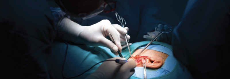
Two surgical approaches are used to correct SSCD, Formeister says. The decision about which is best depends on the patient’s anatomy, age, medical comorbidities, and preferences.
A transmastoid procedure allows surgeons to access the canal through the mastoid bone behind the year. In some cases, this can be performed as a same-day procedure. The middle fossa craniotomy requires a small hole in the skull just above the ear to improve surgeons’ view of and access to the superior canal and requires two nights in the hospital.